Anatomy Rib Cage Muscles / Muscles Of The Rib Cage Labeled Intercostal Muscles Function Area Course Human Click On The Tags Below To Find Other Quizzes On The Same Subject Decorados De Unas : Some muscles of upper body again with neck muscles and deltoids.
Anatomy Rib Cage Muscles / Muscles Of The Rib Cage Labeled Intercostal Muscles Function Area Course Human Click On The Tags Below To Find Other Quizzes On The Same Subject Decorados De Unas : Some muscles of upper body again with neck muscles and deltoids.. As with the muscles that work on the ribcage, these muscles can be used to move the shoulder blades and collar bones to help extend the reach of the arms (downwards, forwards, upwards. Anatomy study anatomy reference art reference anatomy bones body anatomy rib cage anatomy skull anatomy life drawing figure. Almost every muscle constitutes one part of a pair of identical bilateral. Anatomy of a muscle cell. What is superficial to deep?
This is a table of skeletal muscles of the human anatomy. As with the muscles that work on the ribcage, these muscles can be used to move the shoulder blades and collar bones to help extend the reach of the arms (downwards, forwards, upwards. They are curved and flat bones. Smooth muscles are found in the walls of many organs, such as the stomach and in blood vessels. Microscopic anatomy of skeletal muscle.
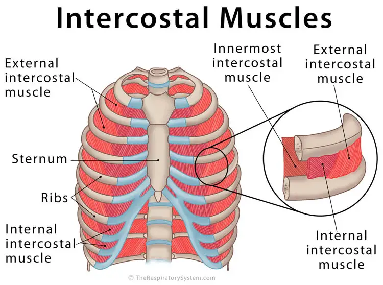
Intercostal Muscles Definition Location Anatomy Functions from www.therespiratorysystem.com Learn anatomy faster and remember everything you learn. Other muscles, like the skeletal muscle that moves the arm, is controlled by the somatic or voluntary nervous system. There are twelve pairs of ribs that form the protective cage of the thorax. What is superficial to deep? Find the best weight lifting exercises that target each muscle or groups of muscles. This muscle is in the middle and has no muscles posterior to it. Anatomy of the muscular system. Cardiac muscles are found in the walls of the heart.
Muscles are named according to their shape, location, or a combination.
Noticing the relationship of the latissimus and the teres ma развернуть. The first drawing showcases the latissimus dorsi muscles at the side of the ribcage. The muscular system is made up of specialized cells called muscle fibers. The muscles of the torso, examined in the previous chapter, include a few that attach directly into the upper arm and help move the humerus at the shoulder joint. Copic fineliner, copic markers, and uniball signo white gel pen on strathmore toned gray paper and some pastels. Molly smith dipcnm, mbant • reviewer: The muscle fibers of the external obliques run diagonally downward and inward from the lower ribs to the pelvis, forming the letter v. This page describes skeletal muscle development, descriptions of cardiac muscle and smooth muscle development can be found in other notes. Each type of muscle tissue in the human body has a unique structure and a specific role. Microscopic anatomy of skeletal muscle. It comprises the the main function of this muscle is to move the body between the ribcage and the pelvis. It impairs full expansion of the ribcage, thus affecting the oxygen content of the blood. Human anatomy for muscle, reproductive, and skeleton.
The first drawing showcases the latissimus dorsi muscles at the side of the ribcage. Smooth muscles are found in the walls of many organs, such as the stomach and in blood vessels. This page describes skeletal muscle development, descriptions of cardiac muscle and smooth muscle development can be found in other notes. You can locate them by putting your hands in your coat pockets. Anatomy of the muscular system.
Back Pain And Slipped Rib from www.spineuniverse.com This page describes skeletal muscle development, descriptions of cardiac muscle and smooth muscle development can be found in other notes. The first drawing showcases the latissimus dorsi muscles at the side of the ribcage. This muscle forms the anterior and lateral abdominal wall. Cardiac muscles are found in the walls of the heart. They are curved and flat bones. Muscles are named according to their shape, location, or a combination. They're shapes that won't change too much between poses. There are around 650 skeletal muscles within the typical human body.
They're shapes that won't change too much between poses.
When the ribcage is fixed contraction results in a posterior pelvic tilt. Their main function is contractibility. Located immediately below the skin) muscles of the body. Each type of muscle tissue in the human body has a unique structure and a specific role. The first drawing showcases the latissimus dorsi muscles at the side of the ribcage. This muscle forms the anterior and lateral abdominal wall. Anatomy of the muscular system. Microscopic anatomy of skeletal muscle. They also contract involuntarily, but have a. Human muscle system, the muscles of the human body that work the skeletal system, that are under voluntary control, and that are concerned with movement, posture, and balance. In this episode, i'll show you how to draw the forms of the rib cage step by step.giveaway! Molly smith dipcnm, mbant • reviewer: Muscles are groups of cells in the body that have the ability to contract and relax.
Microscopic anatomy of skeletal muscle. Their main function is contractibility. Noticing the relationship of the latissimus and the teres ma развернуть. Muscles are named according to their shape, location, or a combination. Anatomy of the muscular system.
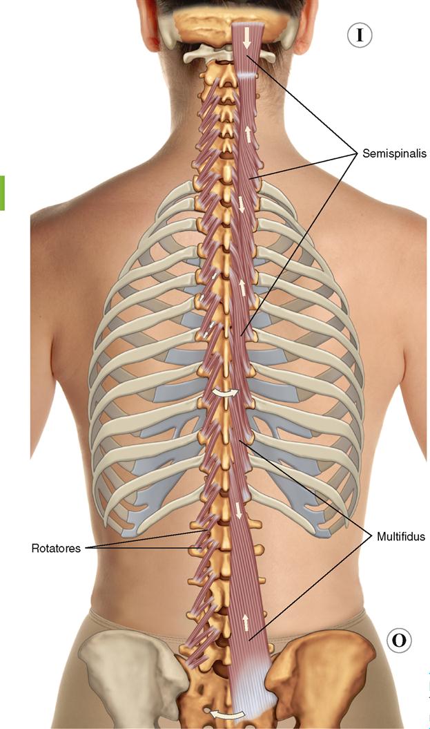
8 Muscles Of The Spine And Rib Cage Musculoskeletal Key from musculoskeletalkey.com Each type of muscle tissue in the human body has a unique structure and a specific role. When the ribcage is fixed contraction results in a posterior pelvic tilt. *completed*if you'd like to win a free membership to the premium. In the muscular system, muscle tissue is categorized into three distinct types: Their main function is contractibility. Dimitrios validated and aligned with popular anatomy textbooks, these muscle cheat sheets are packed with. Anatomy of a muscle cell. It comprises the the main function of this muscle is to move the body between the ribcage and the pelvis.
It should be noted that there are many more.
They also contract involuntarily, but have a. Learn anatomy faster and remember everything you learn. This muscle is in the middle and has no muscles posterior to it. Related posts of muscle anatomy rib cage. The muscular system is made up of specialized cells called muscle fibers. It impairs full expansion of the ribcage, thus affecting the oxygen content of the blood. Microscopic anatomy of skeletal muscle. Muscles are groups of cells in the body that have the ability to contract and relax. They're shapes that won't change too much between poses. The skull, ribcage and pelvic bone are fairly solid and rigid parts of the body (though not always completely rigid). Muscles are named according to their shape, location, or a combination. Human anatomy for muscle, reproductive, and skeleton. Cardiac muscles are found in the walls of the heart.
The muscle fibers of the external obliques run diagonally downward and inward from the lower ribs to the pelvis, forming the letter v anatomy rib cage. This muscle is in the middle and has no muscles posterior to it.


0 Komentar